|
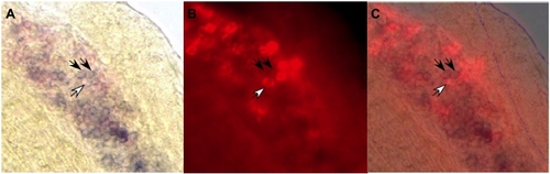
The Mpo+ cells accumulated in Tg(hsp:AML1-ETO) embryos are unlikely to be immature erythroid progenitors. (A) Bright field and (B) fluorescent image of double in situ hybridization of Mpo (purple color, no fluorescence) and βe3 globin (red color, red fluorescence) at 31 hpf. (C) Combined image of both bright field and fluorescence image. The photos show the side view of the embryo close to the end of yolk extension (marked in blue in C). Two filled arrows point to the Mpo+ cells stained purple but have no red fluorecence, suggesting that these cells do not express βe3 globin. By contrast, the cell that expresses βe3 globin does not express Mpo (open arrow).
|