
Fig. 1
- ID
- ZDB-FIG-071223-1
- Publication
- Yabe et al., 2007 - The zebrafish maternal-effect gene cellular atoll encodes the centriolar component sas-6 and defects in its paternal function promote whole genome duplication
- Other Figures
- All Figure Page
- Back to All Figure Page
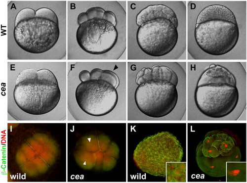
Phenotype of maternally mutant cea embryos. (A–H) Side views of live wild-type (A–D) and mutant (E–H) embryos at the following time points: (A, E) 45 min post fertilization (p.f.), (B, F) 1.25 h p.f., (C, G) 2.25 h p.f., (D, H) 3 h p.f. (corresponding, in wild-type, to the 2-, 8-, 128- and 1000-cell stages, respectively). Maternally mutant cea embryos show a normal first cell division (E) but fail in a fraction of subsequent divisions (F, arrowhead), leading to blastula with irregularly sized blastomeres (G) and largely syncytial embryos (H). (I–L) Animal views of fixed wild-type (I, K) and mutant (J, L) embryos labeled to detect β-catenin (green) and DNA (red). At 1.25 h p.f. (8-cell stage in wild-type; panels I, J), the mutant embryo reveals a failure of DNA and cell division in a fraction of the blastomeres (arrowheads indicate nuclei of cells that did not undergo division). At 3 h p.f. (1000-cell stage in wild-type; panels K, L), mutant embryos exhibit enlarged nuclei (inset in panel L, compare to panel K). |
| Fish: | |
|---|---|
| Observed In: | |
| Stage Range: | 2-cell to 1k-cell |
Reprinted from Developmental Biology, 312(1), Yabe, T., Ge, X., and Pelegri, F., The zebrafish maternal-effect gene cellular atoll encodes the centriolar component sas-6 and defects in its paternal function promote whole genome duplication, 44-60, Copyright (2007) with permission from Elsevier. Full text @ Dev. Biol.