
Fig. 4
- ID
- ZDB-FIG-071120-10
- Publication
- Webb et al., 2007 - Laminin alpha5 is essential for the formation of the zebrafish fins
- Other Figures
- All Figure Page
- Back to All Figure Page
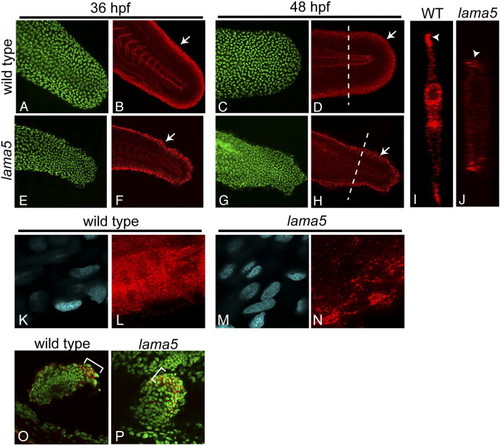
Laminin α5 is essential for basement membrane assembly in the developing fin fold. Immunostaining for laminin (red) and p63 (green) in wild-type (A–D) and lama5 mutants (E–H) shows disruptions in basement membrane along the fin fold margins (arrows) at 36 (B, F) and 48 hpf (D, H). p63 staining marks epidermal cells (A, C, E, G). (I, J) Confocal line scans (transverse sections) at the level of the dotted lines in panels D and H show laminin deposition at the tips of wild-type and mutant fin folds (arrowheads). (K–N) Higher magnification views of laminin staining in wild-type (L) and mutant (N) embryos show aberrations in basal laminae in lama5 mutants. (K, M) DAPI-stained nuclei (blue) appear morphologically normal in the affected region. (O, P) Immunostaining for laminin (red) in the pectoral fins of wild-type (O) and lama5 mutant (P) embryos at 44 hpf. Laminin proteins localize to the margin of the developing pectoral fin and are enriched in the posterior region in both wild-type and mutant embryos (brackets). DAPI-stained nuclei are pseudo-colored green for contrast. In panels A–H and K–N, dorsal is up and anterior is to the left. Pectoral fin views are dorsal with anterior to the left. |
| Fish: | |
|---|---|
| Observed In: | |
| Stage Range: | Prim-25 to Long-pec |
Reprinted from Developmental Biology, 311(2), Webb, A.E., Sanderford, J., Frank, D., Talbot, W.S., Driever, W., and Kimelman, D., Laminin alpha5 is essential for the formation of the zebrafish fins, 369-382, Copyright (2007) with permission from Elsevier. Full text @ Dev. Biol.