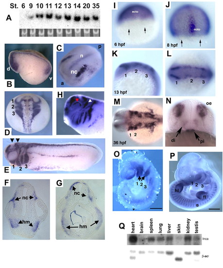
Expression patterns in Xenopus, zebrafish and mouse embryos. (A) Developmental northern blot of Xenopus Inca, with 18S RNA as a loading control. Nieuwkoop-Faber stages are indicated. (B-H) Whole-mount in situ hybridization for Inca in Xenopus embryos at stages 10.5-32. (B) Stage 10.5, sagittal section. d, dorsal. v, ventral. (C) Stage 14, dorsal view of expression in cranial NC (nc) and notochord (n). a, anterior. p, posterior. (D) Stage 19, dorsal view of expression in cranial NC migration streams, labeled as 1-3. (E) Stage 25, lateral view; arrowheads indicate approximate section levels in F and G. NC migration streams into pharyngeal arches 1-3 as indicated. (F,G) Transverse sections of embryo in E show Inca expression in head mesenchyme (hm) and NC (nc). (H) Stage 32, lateral view of the head. White and red arrowheads indicate expression in trigeminal ganglion and eye, respectively. (I-N) Whole-mount in situ hybridization for inca1 in zebrafish embryos. (I) 6 hpf, lateral view, dorsal to the right. Expression is restricted away from the margin (arrows), in future ectoderm (ecto). (J) 8 hpf, dorsal view. noto, notochord. (K,L) 13 hpf, lateral and dorsal views, respectively, of inca1 expression in premigratory NC. Numbers indicate presumptive pharyngeal arches 1-3. (M,N) 36 hpf, dorsal and face-on views, showing expression in the pharyngeal arches (1,2), diencephalon (di), pituitary (pi) and olfactory epithelia (oe). (O,P) Inca expression in mouse embryos at E9.5 (O) and E11.5 (P) in pharyngeal arches (numbered 1-3), somites (s) and fore-limb (fl) and hind-limb buds (hl). (Q) Northern analysis of adult mouse tissue RNAs probed with Inca and beta-actin as control. Scale bars: 500 μm in O; 1 mm in P.
|
