
Fig. 4
- ID
- ZDB-FIG-071005-39
- Publication
- Matthews et al., 2004 - The zebrafish onecut gene hnf-6 functions in an evolutionarily conserved genetic pathway that regulates vertebrate biliary development
- Other Figures
- All Figure Page
- Back to All Figure Page
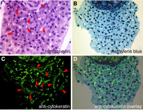
Immunohistochemical detection of zebrafish bile ducts. (A) Histological cross section through the 5-dpf larval liver. Bile ducts are not recognizable in samples processed with standard fixatives (paraformaldehyde; hematoxylin staining). Arrowheads point to vascular sinusoids (clear regions) that contain nucleated red blood cells. (B) Histological cross section through the liver section of a whole-mount specimen (5 dpf) processed for cytokeratin IHC (methylene blue staining). (C) Fluorescent image of section in (B) showing bile ducts and vascular sinusoids (arrowheads). (D) Computer-assisted overlay of (C) on (B). |
Reprinted from Developmental Biology, 274(2), Matthews, R.P., Lorent, K., Russo, P., and Pack, M., The zebrafish onecut gene hnf-6 functions in an evolutionarily conserved genetic pathway that regulates vertebrate biliary development, 245-259, Copyright (2004) with permission from Elsevier. Full text @ Dev. Biol.