
Fig. 5
- ID
- ZDB-FIG-071004-28
- Publication
- Iovine et al., 2005 - Mutations in connexin43 (GJA1) perturb bone growth in zebrafish fins
- Other Figures
- All Figure Page
- Back to All Figure Page
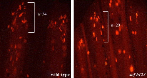
Cell proliferation during ontogenetic growth in wild-type and sofb123 caudal fins. Wild-type and sofb123 fish were labeled with BrdU for 6 h. BrdU-positive cells are labeled in red. The bracket indicates the region from which cells were counted, n indicates the number of BrdU-positive cells in this particular fin ray (note: all BrdU-positive cells are not in this focal plane). Left: wild-type fin ray showing BrdU-labeled cells. On average, 32 ± 5 cells are labeled in the third fin ray from either lobe (n = 10 fins). Right: sofb123 fin ray showing BrdU-labeled cells. On average, 21 ± 3 cells are labeled in the third fin ray from either lobe (n = 10 fins). |
Reprinted from Developmental Biology, 278(1), Iovine, M.K., Higgins, E.P., Hindes, A., Coblitz, B., and Johnson, S.L., Mutations in connexin43 (GJA1) perturb bone growth in zebrafish fins, 208-219, Copyright (2005) with permission from Elsevier. Full text @ Dev. Biol.