
Fig. 9
- ID
- ZDB-FIG-070917-69
- Publication
- Rojas-Munoz et al., 2005 - chokh/rx3 specifies the retinal pigment epithelium fate independently of eye morphogenesis
- Other Figures
- All Figure Page
- Back to All Figure Page
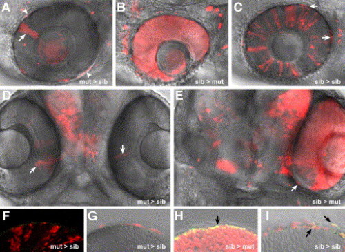
chk/rx3 specifies the prospective RPE. Single confocal images of mosaic eyes generated by transplantation of rhodamine-labeled cells at the late blastula stage. chks mutant cells transplanted into wt embryos form small clones of NR (arrows in panels A and D) but are completely excluded from the RPE. Some cells are false positives as concluded by the analysis of the complete Z stack (asterisks in panel A). Importantly, even donor cells in close proximity to the eye can be precisely considered as excluded from the RPE (arrowheads in panel A). Conversely, wt cells transplanted into chks mutant embryos form NR and RPE, while the host cells are always excluded from the RPE (B, E). Notably, even in a fully rescued eye with mosaic NR, the RPE is of wt origin (arrow in E). Donor cells randomly contribute to the RPE in control transplanted embryos (arrows in panel C). Specific staining of the RPE with anti-RPE65 (green in panels F–I). Overlapping signal is observed in the RPE only in sib > mut (H) or sib > sib (I) mosaics (black arrows). The images correspond to 70 hpf larvae. The orientation of the chimeras is dorsal up, anterior to the left (A, B, C) or ventral view, anterior upward (D, E). |
Reprinted from Developmental Biology, 288(2), Rojas-Munoz, A., Dahm, R., and Nüsslein-Volhard, C., chokh/rx3 specifies the retinal pigment epithelium fate independently of eye morphogenesis, 348-362, Copyright (2005) with permission from Elsevier. Full text @ Dev. Biol.