
Fig. 3
- ID
- ZDB-FIG-070914-14
- Publication
- Theusch et al., 2006 - Separate pathways of RNA recruitment lead to the compartmentalization of the zebrafish germ plasm
- Other Figures
- All Figure Page
- Back to All Figure Page
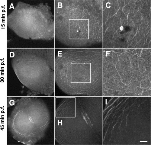
An f-actin network undergoes arrangements that are similar to those of the RNA-bound filaments. Animal views of embryos fixed at progressively older stages of development (A–C: 15 min p.f.; D–F: 30 min. p.f.; G–I: 45 min p.f.) and labeled with fluorescent phalloidin. Panels A–C, D–F, and G–I are sets of progressively higher magnifications of the same embryo (left column: overview; middle and left columns: square in the middle panel is shown in the right panel at a higher magnification). (A–C) Initially f-actin forms a network of randomly aligned cables. (D–F) As the first cell cycle proceeds, f-actin begins to move towards the periphery (note clearing of f-actin in the center of the blastodisc) and circumferential alignment (note alignment of outer cables). (G–I) These effects become more pronounced at later stages of development. The accumulation of f-actin in panels B, C highlights the second polar body, which forms after egg activation (Dekens et al., 2003). The accumulation of f-actin at the center of the blastodisc in panels G, H corresponds to f-actin enrichments that typically occur at the site of furrow formation (Kishimoto et al., 2004; A. Menzie and F.P., unpublished). Scale bar in panel I represents 100 μm (left column), 50 μm (middle column), and 16 μm (right column). |
Reprinted from Developmental Biology, 292(1), Theusch, E.V., Brown, K.J., and Pelegri, F., Separate pathways of RNA recruitment lead to the compartmentalization of the zebrafish germ plasm, 129-141, Copyright (2006) with permission from Elsevier. Full text @ Dev. Biol.