
Fig. 1
- ID
- ZDB-FIG-070911-81
- Publication
- Tanaka et al., 2007 - Novel mutations affecting axon guidance in zebrafish and a role for plexin signalling in the guidance of trigeminal and facial nerve axons
- Other Figures
- All Figure Page
- Back to All Figure Page
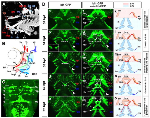
The stepwise axonal pathfinding of the Vp and VII motoneurons. The cranial axons and jaw muscles were visualised using the Isl1-GFP and α-actin-GFP double-transgenic strain. (A,B) The jaw region of a 72-hpf zebrafish embryo. Anterior, left. (B) Schematic of the projection pattern of the Va, Vp and VII motoneurons. Thick black and white arrows indicate the points at which these motor axons separate from the common pathways. (C) Dorsal view of the hindbrain of a 72-hpf Isl1-GFP transgenic zebrafish embryo. Anterior, top. The Va, Vp and VII motor nuclei are located in r2, r3 and r6, respectively. (Da-o) The stepwise outgrowth of the Vp (red) and VII (blue) motoneurons from 52-72 hpf. Ventral view; anterior, top. Broken lines indicate the boundaries between BA1 and BA2 (b-e,g-j). Arrowheads indicate the points at which the growth cones of the VII motoneurons stalled at 54-58 hpf (b-d,g-i). Va, anterior trigeminal motoneurons; Vp, posterior trigeminal motoneurons; VII, facial motoneurons; fs, facial sensory ganglion; tg, trigeminal sensory ganglion; BA1, BA2, first and second branchial arches; r, rhombomere; ah, adductor hyomandibulae; am, adductor mandibulae; ao, adductor operculi; do, dilatator operculi; hh, hyohyal; ih, interhyal; ima, intermandibularis anterior; imp, intermandibularis posterior; lap, levator arcus palatini; sh, sternohyoideus. |