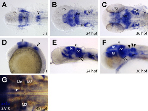
Developmental expression of irx1a in the zebrafish brain. A-F: Dorsal view of flat-mounted (A-C) and a lateral view (D-F) of the zebrafish brain region. A,D: irx1a is expressed in the prospective midbrain and the anterior hindbrain regions but not in the midbrain-hindbrain boundary at the five-somite stage (5 s). Note that irx1a is also weakly expressed in the posterior hindbrain region (open arrowheads). B,E: irx1a is expressed in the tectum, cerebellum, rostral hindbrain region, and acoustic ganglion at 24 hpf. C,F: By 36 hpf, expression of irx1a can be detected in the lateral line. In the hindbrain, irx1a expression is extended to the caudal part of hindbrain and in the commissural neurons (black arrowheads). G: Dorsal view of the hindbrain region of flat-mounted 48 hours postfertilization (hpf) embryo; double in situ hybridizaton and immunostaining staining of irx1a (dark blue) and 3A10 (brown). irx1a is not expressed in the reticulospinal neurons, and the Mauthner axons (white arrow) lie just dorsal to the ventral irx1a expression domain. Asterisks (*) indicate the position of midbrain-hindbrain boundary. ac, acoustic ganglion; ce, cerebellum; ey, eye; la, lateral line; Mn, Mauthner neuron; M2, Mi2cm; M3, Mi3cm; ov, otic vesicle; r1, rhombomere 1; te, tectum; tg, tegmentum.
|
