
Fig. 1
- ID
- ZDB-FIG-070911-15
- Publication
- Moore et al., 2007 - A role for the Myoblast city homologues Dock1 and Dock5 and the adaptor proteins Crk and Crk-like in zebrafish myoblast fusion
- Other Figures
- All Figure Page
- Back to All Figure Page
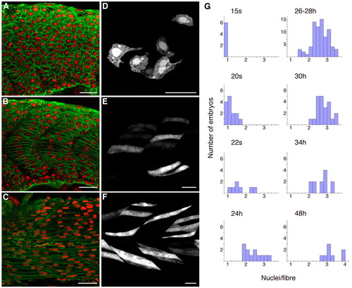
Wild-type fusion of fast-twitch myoblasts. (A-C) Mid-trunk; β-catenin (green) and DAPI (red). (A) At 18 somites; optical section mid-way between the midline and the lateral surface, through fast precursors, showing unfused small rounded cells. (B) At 18 somites; optical section through medial adaxial cells, the slow-fibre precursors, which have elongated. (C) At 26 hpf; optical section through fast muscle fibres, showing multinucleate fused cells. (D-F) mylz2:GFP transgene expression in isolated cells. GFP labelling is found almost exclusively in fast-twitch muscle fibres, the few slow fibres that express the transgene being distinguishable by their shape and more-advanced differentiative state. (D) At 17 somites, showing small rounded mostly mononucleate cells with projections. (E) At 20 somites, showing that fusion has started to occur. Some cells are multinucleate. (F) At 26 hpf, showing elongated multinucleate fibres. (G) Quantitative data showing distributions of mean nuclei/fibre for individual wild-type embryos fixed at progressive embryonic stages, from 15 somites (15s) to 48 hpf (48h). Scale bars: 25 μm. |