
Fig. 2
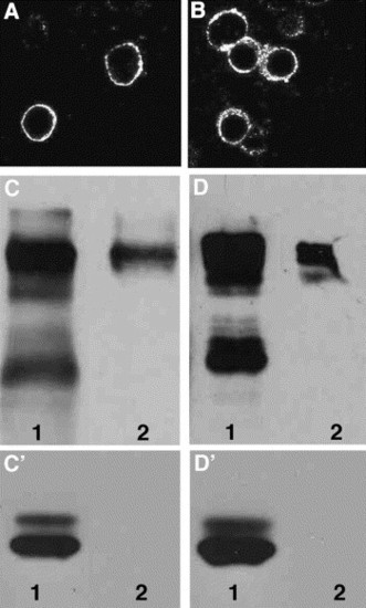
Protein localization of Robo3 isoforms in transiently transfected mammalian cells. myc-tagged (A) robo3var1 and (B) robo3var2 isoforms driven by a CMV promoter were transfected into 293T cells. Protein expression was detected by immunofluorescence using a rhodamine conjugated anti-Myc antibody. (C) 293T cells were transfected with myc-tagged robo3var1 or (D) robo3var2 followed by biotinylation of surface proteins. Lane 1 (C–D′) is the flowthrough and reflects cytoplasmic proteins or proteins that did not bind to avidin. Lane 2 (C–D′) is surface proteins that are biotinylated and bound to avidin. Proteins in C, D are recognized by an anti-myc antibody. (C′, D′) The same blot was stripped and reprobed with anti-actin antibody. Due to lower expression of Robo3var2 in transfected cells, Lanes D 1,2 were exposed longer compared to lanes C 1,2. Lanes C′ and D′ were exposed for the same amount of time reflecting similar amounts of total protein. |
Reprinted from Mechanisms of Development, 122(10), Challa, A.K., McWhorter, M.L., Wang, C., Seeger, M.A., and Beattie, C.E., Robo3 isoforms have distinct roles during zebrafish development, 1073-1086, Copyright (2005) with permission from Elsevier. Full text @ Mech. Dev.