
Fig. 7
- ID
- ZDB-FIG-070821-18
- Publication
- Shimizu et al., 2005 - E-cadherin is required for gastrulation cell movements in zebrafish
- Other Figures
- All Figure Page
- Back to All Figure Page
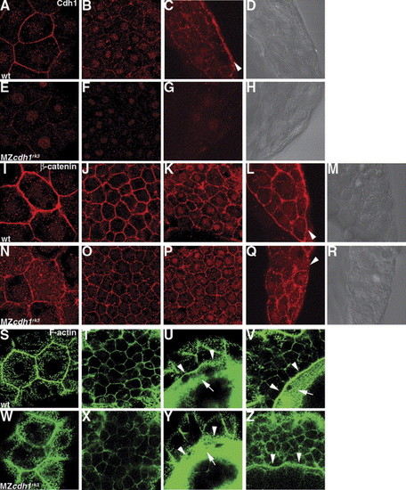
Cell adhesion within DCs and microfilament formation at the margin of the EVL and DCs are maintained in the cdh1 morphant embryos. Wild-type (wt) and MZcdh1rk3 mutant embryos were fixed at 8.5 hpf (80% epiboly for wild-type), and stained with an anti-Cdh1 antibody (A–H), anti-β-catenin antibody (I–R), or Alexa Fluor 488-phalloidin (S–Z). Immunostaining was visualized by Alexa Fluor 568-conjugated secondary antibody (A–R). (A–D, I–M, S–V) Wild-type embryos. (E–H, N–R, W–Z) MZcdh1rk3 mutant embryos. Confocal microscopic optical sections in the animal pole region at the level of the enveloping layer (A, E, I, N, S, W) and deep cells (B, F, J, O, T, X). Confocal sections in the lateral blastoderm margin (C, D, G, H, K–M, P–R), cross sections of the dissected embryos (C, D, G, H, L, M, Q, R), and optical sections at the level of the deep cells (K, P). (D, H, M, R) DIC images of (C, G, L, Q). Confocal optical section of the epiboly margin at the level of the EVL (U, Y) and deep cells (V, Z). Cdh1 protein was strongly detected in the contact surface between the EVL and the DCs (arrowhead in C). Accumulation of β-catenin was detected on the contact surface between the YSL and the DCs in the wild-type embryos but was severely decreased in the MZcdh1rk3 mutant embryos (arrowheads in L, Q). The ring-like F-actin-based structures were detected in the edges of the EVL (arrowheads, U, Y) and the edge of the DCs (arrowheads in V, Z), and punctate actin bands were detected in the E-YSL (arrows in U, V, Y), adjacent to the edges of the EVL and the DCs in the wild-type embryos, and to the edge of the EVL in the MZcdh1rk3 mutant embryos. |
| Antibody: | |
|---|---|
| Fish: | |
| Anatomical Terms: | |
| Stage: | 75%-epiboly |
Reprinted from Mechanisms of Development, 122(6), Shimizu, T., Yabe, T., Muraoka, O., Yonemura, S., Aramaki, S., Hatta, K., Bae, Y.K., Nojima, H., and Hibi, M., E-cadherin is required for gastrulation cell movements in zebrafish, 747-763, Copyright (2005) with permission from Elsevier. Full text @ Mech. Dev.