
Fig. 5
- ID
- ZDB-FIG-070821-12
- Publication
- Shi et al., 2005 - Zebrafish pitx3 is necessary for normal lens and retinal development
- Other Figures
- All Figure Page
- Back to All Figure Page
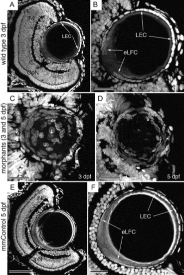
Morphology and spatial organization of the lens nuclei. Propidium iodide-stained wild type (A, B), morphant (C, D) and mismatch control (E, F) frozen eye sections. At 3 dpf, the normal retinal laminar arrangement (A) and lens epithelial cell organization (B) is shown. The epithelial cells covering the distal aspect of the lens (LEC) and the proximal elongating fiber cells (eLFC) are indicated by arrows (A, B). The 3 dpf morphant lens retains the central fiber cell nuclei (C) and the epithelial cell population exhibits enlarged and irregularly shaped nuclei and multilayering at 3 and 5 dpf (C, D). The mismatch control retina and lens (E, F) are indistinguishable from wild type (A, B). Abbreviations: PL, photoreceptor layer; INL, inner nuclear layer; GCL, ganglion cell layer; LEC, lens epithelial cells; eLFC, elongating lens fiber cells. Scale bars in A, B, E and F represent 50 μm, while the scale bars in C and D are 20 μm. |
Reprinted from Mechanisms of Development, 122(4), Shi, X., Bosenko, D.V., Zinkevich, N.S., Foley, S., Hyde, D.R., Semina, E.V., and Vihtelic, T.S., Zebrafish pitx3 is necessary for normal lens and retinal development, 513-527, Copyright (2005) with permission from Elsevier. Full text @ Mech. Dev.