
Fig. 5
- ID
- ZDB-FIG-070802-7
- Publication
- Tessmar-Raible et al., 2007 - Conserved sensory-neurosecretory cell types in annelid and fish forebrain: insights into hypothalamus evolution
- Other Figures
- All Figure Page
- Back to All Figure Page
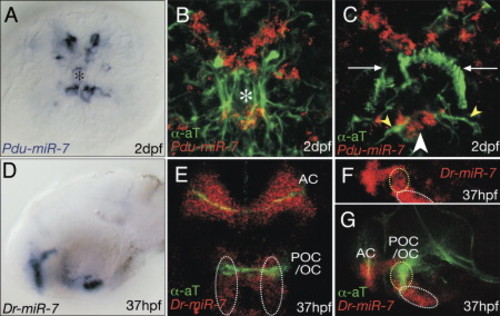
miR-7 Demarcates Specific Groups of Cells in the Medial Forebrain of Platynereis and Zebrafish. Expression of miR-7 in relation to the axonal scaffold (green) in (A–C) Platynereis and (D–G) zebrafish. (A–C) Apical views, ventral to the bottom. White arrows: position of large multiciliated crescent cell as landmark for orientation; yellow arrowheads: microtubule-rich dendrite; asterisks: position of neurosecretory forebrain plexus. (D, F, and G) Lateral views, anterior to the left, (E) ventral view, anterior: top. Yellow and white circles outlines the same groups of cells in Figures 3M, 4J, and 5E–5G. AC, anterior commissure; POC/OC, postoptic commissure/optic chiasm. |
| Gene: | |
|---|---|
| Fish: | |
| Anatomical Term: | |
| Stage: | Prim-25 |
Reprinted from Cell, 129(7), Tessmar-Raible, K., Raible, F., Christodoulou, F., Guy, K., Rembold, M., Hausen, H., and Arendt, D., Conserved sensory-neurosecretory cell types in annelid and fish forebrain: insights into hypothalamus evolution, 1389-1400, Copyright (2007) with permission from Elsevier. Full text @ Cell