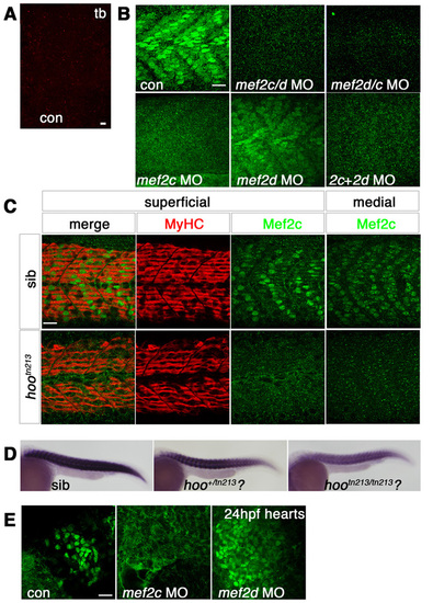
Morpholino knockdown of mef2c. Immunofluorescent detection of anti-Mef2c antibody (A, red; B-D, green) or slow MyHC (C, red) in skeletal muscle viewed in dorsal (A, presomitic mesoderm, anterior to top) or lateral (B,C, anterior to left, dorsal to top) flatmount. (A) Confocal stack showing unstained adaxial cells in presomitic mesoderm at tailbud stage. (B) At 24 hpf, confocal stacks reveal that Mef2c immunoreactivity is predominantly nuclear in both fast and slow muscle fibres. mef2c MO ablates nuclear signal, whereas mef2d MO has little effect. mef2c/d, mef2d/c and mef2c + mef2d MOs also ablate Mef2c immunoreactivity. (C) Single confocal slices of 24-hpf embryos from a cross of hoover heterozygotes. Mef2c immunoreactivity is ablated in superficial slow and medial fast fibres of 13/48 (27%) hootn213 mutant embryos, but slow muscle is unaffected. (D) mef2c mRNA is reduced in putative hootn213 mutants (21/67, 31%) and partially reduced in heterozygotes (27/67, 40%). (E) At 24 hpf, Mef2c immunoreactivity is predominantly nuclear in cardiomyocytes. mef2c MO ablates nuclear signal, whereas mef2d MO has no effect. Note that mef2c-specific MO greatly reduced anti-Mef2c stain in heart and muscle and anti-Mef2 in heart without diminishing muscle anti-Mef2 signal or cognate mRNA (compare to Fig. S1A-C). Scale bars: 20 μm.
|
