
Fig. 5
- ID
- ZDB-FIG-070613-15
- Publication
- Lillesaar et al., 2007 - The serotonergic phenotype is acquired by converging genetic mechanisms within the zebrafish central nervous system
- Other Figures
- All Figure Page
- Back to All Figure Page
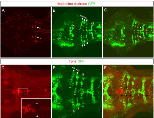
Location of the gap separating the anterior and posterior pet1-positive raphe precursors in relation to green fluorescent protein (GFP) -positive cell clusters in the isl1:gfp transgenic line. Photomicrographs are confocal optical projections of dorsal views at hindbrain levels, anterior left. A: Mauthner neurons (arrows), located in rhombomere (r) 4, were labeled by retrograde tracing in 6 days postfertilization (dpf) isl1:gfp transgenic larvae using rhodamine dextran. B,C: Thereby, the spatial distribution of GFP-expressing cells (B, arrowheads) in relation to rhombomeres was determined (C, overlay of A and B). C: Note that the Mauthner neurons overlap with a cluster of GFP-positive cells (*) just posterior to the trigeminal nuclei (Va and Vp, arrows). D: The anterior and posterior clusters of serotonergic precursors were identified using an antibody against tryptophan hydroxylase (Tph) 2. The inset shows a higher magnification of the boxed area, and white arrowheads in the inset indicate the gap between the anterior and posterior Tph-positive clusters (optical section). E,F: Note, in F (overlay of D and E) that this gap overlaps with Vp, in r3 (black arrowheads), rather than with Mauthner neurons, in r4. Thus, the anterior Tph cluster spans r1-2, and the posterior cluster spans r4 and beyond. |