
FIGURE
Fig. 6
- ID
- ZDB-FIG-070418-5
- Publication
- Ott et al., 2007 - Comparative analysis of splice form-specific expression of LIM Kinases during zebrafish development
- Other Figures
- All Figure Page
- Back to All Figure Page
Fig. 6
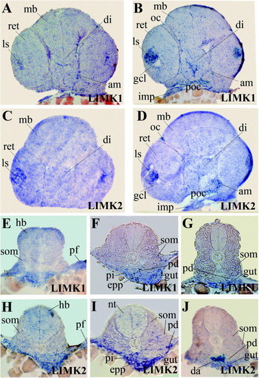
Shown are sections of whole mount in situ hybridizations of long pec stage embryos probed for LIMK1 and LIMK2, respectively. Abbreviations used are: adductor mandibulae (am), diencephalon (di), dorsal aorta (da), endoderm (e), exocrine pancreatic progenitor (epp), ganglion cell layer (gcl), hindbrain (hb), intermandibularis posterior (imp), lens (ls), midbrain (mb), notochord (nt), optic chiasm (oc), pectoral fin buds (pf), posterior chiasm (poc), pronephric duct (pd), somites (som), retina (ret). |
Expression Data
| Genes: | |
|---|---|
| Fish: | |
| Anatomical Terms: | |
| Stage: | Long-pec |
Expression Detail
Antibody Labeling
Phenotype Data
Phenotype Detail
Acknowledgments
This image is the copyrighted work of the attributed author or publisher, and
ZFIN has permission only to display this image to its users.
Additional permissions should be obtained from the applicable author or publisher of the image.
Reprinted from Gene expression patterns : GEP, 7(5), Ott, E.B., Te Velthuis, A.J., and Bagowski, C.P., Comparative analysis of splice form-specific expression of LIM Kinases during zebrafish development, 620-629, Copyright (2007) with permission from Elsevier. Full text @ Gene Expr. Patterns