
Fig. 3
- ID
- ZDB-FIG-070413-21
- Publication
- Hendricks et al., 2007 - Electroporation-based methods for in vivo, whole mount and primary culture analysis of zebrafish brain development
- Other Figures
- All Figure Page
- Back to All Figure Page
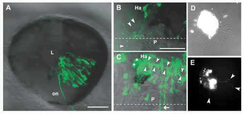
Analysis of transfected neurons in vivo and in vitro. (a) The eye of a 2 dpf embryo electroporated with pHuC:GAL4/pUAS:dnRyk-EGFP. Cells in multiple retinal layers are transfected in a distinct segment according to electrode positioning and injection site. Retinal ganglion cell axons are visible in the optic nerve (on). (b) A habenular (Ha) neuron contransfected with pHuC:GAL4/pUAS:dnEphB3-EGFP shows ectopic processes branching over the medial epithalamus, including the pineal organ (P). Extracellular exosome-like vesicles (arrowheads) are visible around the soma and processes. (c) Two habenular neurons (asterisks) expressing EGFP show the normal ventro-posterior projection into the fasciculus retroflexus (arrowheads). Commissural axons (arrow) are not derived from the habenula. (d) Bright field phase contrast and (e) fluorescence images of a 2 dpf forebrain explant from an embryo electroporated with pHuC:GAL4/pUAS:EGFP after 12 hours in culture. EGFP positive neurons and axons (arrowheads) can be tracked over time. Anterior is to the left in (a) (lateral), (b,c) (dorsal). Dashed lines indicate the midline. Scale bars = 50 μm. L, lens. |