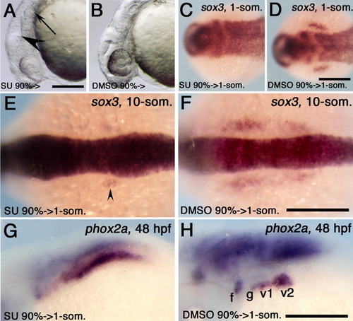
Dependence of the development of the epibranchial placode on the fibroblast growth factor (FGF) signal. A,B: Zebrafish embryo treated with SU5402 (A) and dimethyl sulfoxide (DMSO, B) from 90% epiboly to 26 hours postfertilization (hpf). The disrupted isthmus is shown with a large arrowhead, and an arrow indicates the reduced otic vesicle. C,D:sox3 expression in the head of the one-somite stage embryos treated with SU5402 (C) or DMSO (D) from 90% epiboly stage to the one-somite stage. E-H:sox3 expression at the 10-somite stage (E,F) and phox2a expression at 48 hpf (G,H) were examined in embryos treated with SU5402 (E,G) or DMSO (F,H) from 90% epiboly to the one-somite stage. The small arrowhead indicates residual posterior sox3 expression at this stage. A,B: Lateral views, with anterior to the left and dorsal to the top. C-H: Dorsal views (C-F) or dorsolateral views (G,H), with anterior to the left. The stage of the SU5402 treatment is indicated at the bottom left in each panel. Genes and stages examined are shown at the top right in each panel. f, facial ganglion; g, glossopharyngeal ganglion; v1, v2, vagal ganglion. Scale bar = 200 μm.
|
