
FIGURE
Fig. 6
- ID
- ZDB-FIG-070307-19
- Publication
- Rothschild et al., 2007 - Differential expression of CaMK-II genes during early zebrafish embryogenesis
- Other Figures
- All Figure Page
- Back to All Figure Page
Fig. 6
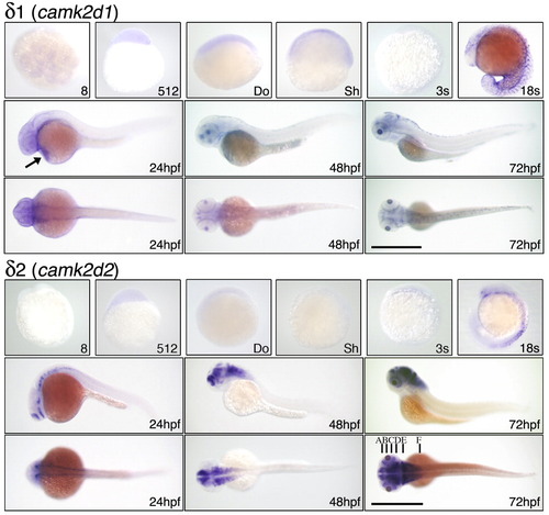
In situ localization of δ CaMK-II mRNAs. CaMK-II expression was assessed by in situ hybridization with a probe for camk2d1 and camk2d2 at the indicated stages as in Figure 4. The 8-cell stage is an animal poll view; others are lateral except for dorsal views at 24, 48, and 72hpf. Arrows in camk2d1 at 24hpf indicates hatching gland. Letters locate cross-sections for Figure 7. Scale bar = 1 mm. |
Expression Data
| Genes: | |
|---|---|
| Fish: | |
| Anatomical Terms: | |
| Stage Range: | 8-cell to Protruding-mouth |
Expression Detail
Antibody Labeling
Phenotype Data
Phenotype Detail
Acknowledgments
This image is the copyrighted work of the attributed author or publisher, and
ZFIN has permission only to display this image to its users.
Additional permissions should be obtained from the applicable author or publisher of the image.
Full text @ Dev. Dyn.