
Fig. 7
- ID
- ZDB-FIG-070228-8
- Publication
- Murayama et al., 2006 - Tracing Hematopoietic Precursor Migration to Successive Hematopoietic Organs during Zebrafish Development
- Other Figures
- All Figure Page
- Back to All Figure Page
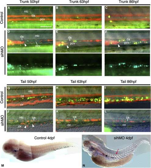
The Seeding of Caudal Hematopoiesis Requires Blood Circulation(A–L) Real-time follow-up of representative control and silent heart morphant [CD41-gfp; gata1-dsred] embryo, at the trunk (A–F) and tail (G–L) level; green+red fluorescence overlaid on DIC images (lateral views, rostral left). In the control embryo, the flow of circulating DsRed+ primitive erythrocytes delineates blood circulation; GFP+ cells develops in the tail (G–I), barely in the trunk (A–C). In the silent heart morphant, many of the noncirculating primitive erythroblasts are gathered in a big mass in the rostral trunk (an arrowhead indicates its caudal border) (D–F); GFP+ cells develop in the trunk ([D]–[F], lower panels show green channel alone) and not in the tail (J–L), despite the presence of a normal CV plexus at 2 dpf ([J], arrowheads). Scale bar represents 100 μm. (M and N) In situ hybridization for c-myb in control and sih morphant embryos. |
| Gene: | |
|---|---|
| Fish: | |
| Knockdown Reagent: | |
| Anatomical Terms: | |
| Stage: | Day 4 |
| Fish: | |
|---|---|
| Knockdown Reagent: | |
| Observed In: | |
| Stage: | Day 4 |
Reprinted from Immunity, 25(6), Murayama, E., Kissa, K., Zapata, A., Mordelet, E., Briolat, V., Lin, H.F., Handin, R.I., and Herbomel, P., Tracing Hematopoietic Precursor Migration to Successive Hematopoietic Organs during Zebrafish Development, 963-975, Copyright (2006) with permission from Elsevier. Full text @ Immunity