|
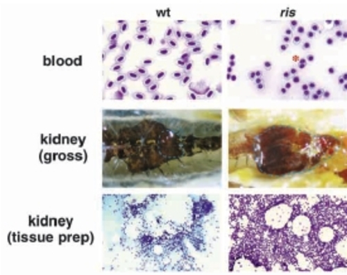
Adult ris blood and marrow. Peripheral blood and kidney collected from wild-type and ris adults. Blood smear and kidney tissue preparation were Wright-Giemsa stained, and whole kidneys are shown in situ, situated in the retroperitoneal cavity of the adult animal (organ margin demarcated with broken lines). Mutant erythrocytes are spherical or tear-drop shaped, with round nuclei. More bi-nucleated cells are seen in the mutant (asterisk). In contrast, the wild-type red cells and nuclei are elliptical. Mutant kidney tissue is dramatically enlarged compared with wild type, with increased cellularity. Blood, gross kidney and tissue kidney are shown at 20x, 4x and 4x, respectively.
|