
FIGURE
Fig. 7
- ID
- ZDB-FIG-061227-25
- Publication
- Hu et al., 2006 - Egr1 gene knockdown affects embryonic ocular development in zebrafish
- Other Figures
- All Figure Page
- Back to All Figure Page
Fig. 7
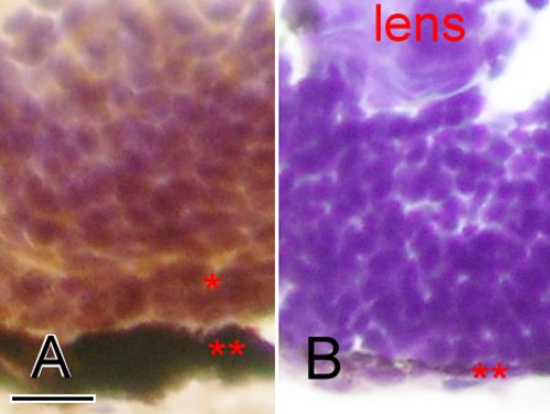
Cryosection and immunostaining of the wildtype and Egr1 morphants. Cryosections of zebrafish eyes at 72 h postfertilization with zpr-1 immunostaining for photoreceptor cells. The wildtype (A) has markedly more labeled retinal cells in the outer nuclear layer (*) than the Grade-3 Egr1 morphant (B). The retinal pigmented epithelial layer (**) is also much thinner in the morphant. The scale bar represents 10 μm in photo A, and is applicable to photo B. |
Expression Data
Expression Detail
Antibody Labeling
Phenotype Data
Phenotype Detail
Acknowledgments
This image is the copyrighted work of the attributed author or publisher, and
ZFIN has permission only to display this image to its users.
Additional permissions should be obtained from the applicable author or publisher of the image.