
Fig. 3
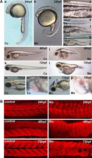
Effects of microinjection of cypher gene-specific morpholinos on zebrafish development. (A) Shown are stereoscope pictures and DIC microscopy pictures of embryos at different stages of development. The magnifications shown are in each case comparable between pictures of control and cypher-MO injected embryos but not for the different stages of embryos. The embryos were injected at the one-cell stage with either a control morpholino or the cypher specific morpholinos (2 ng embryo). a, c, e, g, i, k, m, and n show the control injected and b, d, f, h, j, l, o, and p correspond to the cypher-MO injected embryos. In a and b, embryos at 24 h of development are shown with (a) being the control and (b) the cypher-MO injected embryo. The picture in b of the cypher-MO injected embryo is focused on the somites and the notochord which is present in these embryos is not well visible. The notochord also in the later stages is malformed in cypher-MO injected embryos (d, f, h). Shown in c (control) and d (cypher-MO) are close ups of the region of the first 13 somites of embryos at 48 h of development. The somites of the tail region of 48 hpf embryos are shown in e (coer-MO injected embryos (d, f, h). Shown in c (control) and d (cypher-MO) are close ups of the region of the first 13 somites of embryntrol) and f (cypher-MO). g (control) and h (cypher-MO), the tail regions of 72 hpf embryos are shown. In i and j, embryos at 48 hpf and (k) and (l) at 72 hpf are shown. m and n show the head and heart of 72 hpf embryos of the control injected group whereas o and p show the cypher-MO injected group with enlarged pericardium. (B) Myotome boundaries in control and cypher-MO injected embryos. We used red phalloidin staining to detect F-actin which is present in all muscle cells. Shown on the left are control injected embryos and on the right cypher-MO injected embryos representing the severe phenotype. Time points used were 24 hpf, 48 hpf, and 72 hpf. The loss of organization and of myotome boundaries increases over time. (C) Somite staining in control and cypher-MO injected embryos. Embryos were injected with control (a-c) or cypher-MO (d-f) and fixed at the 15-somite stage. In situ hybridization for vmhc expression (-f) was performed and pictures from the dorsal view were taken. There are severe (f) and less severe deformed embryos (d, e) found. In g and h, in situ hybridization expression for myoD is shown, for a control injec |
| Fish: | |
|---|---|
| Knockdown Reagent: | |
| Observed In: | |
| Stage Range: | Prim-5 to Protruding-mouth |
Reprinted from Developmental Biology, 299(2), van der Meer, D.L., Marques, I.J., Leito, J.T., Besser, J., Bakkers, J., Schoonheere, E., and Bagowski, C.P., Zebrafish cypher is important for somite formation and heart development, 356-372, Copyright (2006) with permission from Elsevier. Full text @ Dev. Biol.