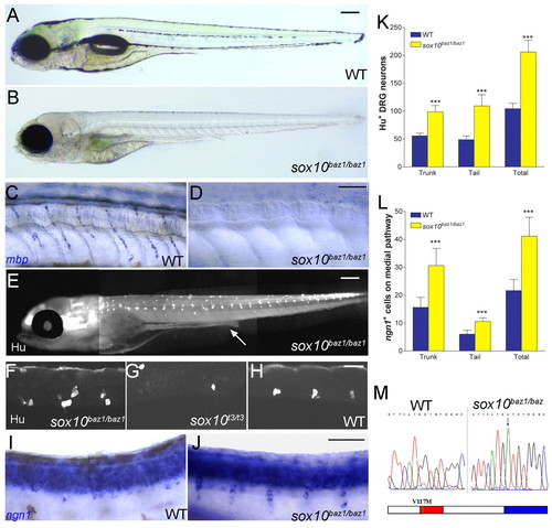
sox10baz1 mutants show a unique, neurogenic DRG phenotype. (A-H) 5 dpf sox10baz1/baz1 showed severe reductions in NC-derived melanophores (B) and mbp+ Schwann cells of spinal nerves (D) compared with wild-type siblings (A,C). Note similar strength of these phenotypes to sox10m618/m618 (see Fig. 2C-D). In contrast, Hu+ DRG sensory neurons were more abundant in sox10baz1/baz1 embryos (E, low power; F, higher confocal magnification view of three segments of tail) than in wild type (H), and thus much more than in typical strong sox10 alleles (G). (I,J) sox10baz1/baz1 embryos similarly displayed an increased number of ngn1+ cells on the medial migration pathway at 36 hpf (J) compared with wild-type (I). (K,L) Quantitation of these Hu+ (K) and ngn1+ (L) DRG-associated cells in sox10baz1/baz1 and siblings (***P<0.0001, Student's t-test; n=10 and n=15 embryos counted for each data set, respectively). (M) Sequencing identified molecular lesion in sox10baz1 as a G to A transition at cDNA position 724 (upper panel), producing a Valine to Methionine substitution at amino acid 117 within the Sox10 protein (lower panel). DNA binding domain (red) and transactivation domain (blue) are indicated. Scale bars: 200 μm in A,B,E; 50 μm in C,D,F-J.
|
