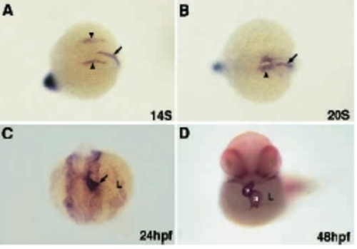
Cardiac morphogenesis in zebrafish. The cardiac precursors are in the ventral margin at the onset of gastrulation in zebrafish. (A) These ventral-derived cardiac precursors migrate toward the animal axis and form bilateral heart primordia on either side of the embryo (arrowhead), flanking the prechordal plate and notochord (arrow) at the 14-somite stage. (B) The bilateral heart tubes then fuse at the midline (arrowhead), anterior to the notochord (arrow). (C) At 24 hours post fertilization (hpf), the primitive heart ‘jogs’ to the left (arrow points to the prospective atrium). The heart then gradually returns to the midline. (D) At 48 hpf, the ventricle of the midline heart loops to the right. The bilateral heart tubes and primitive heart are labeled with Nkx2.5 in A, B and C, and MF20 in D. Notochord precursors are labeled with Brachyury in A and B. (A,B) Dorsallateral view, anterior to the left. (C) Dorsal view, anterior to the bottom of the panel. (D) A ventral view. a, atrium; v, ventricle; L, embryo left.
|
