
Fig. 4
- ID
- ZDB-FIG-061121-14
- Publication
- Lyman Gingerich et al., 2006 - Analysis of axis induction mutant embryos reveals morphogenetic events associated with zebrafish yolk extension formation
- Other Figures
- All Figure Page
- Back to All Figure Page
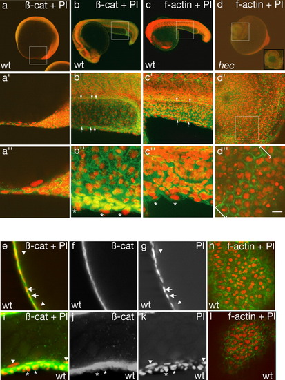
β-Catenin and f-actin localization reveal the presence of an extra layer of cells overlying the enveloping layer (EVL) in the yolk extension region. a,b,e-g,i-k: β-Catenin and propidium iodide labeling of a wild-type embryo. c,d,h,l: f-Actin and propidium iodide labeling of wild-type and hec mutant embryos. a′-d′,a″-d″: Progressively higher magnifications of a-d. Nuclei in the wild-type yolk extension tend to be aligned in rows in a dorsal to ventral direction (white arrows in b′, c′). White asterisks in b″ and c″ denote protruding cells. Site of constriction in radialized hec mutant embryos is shown by brackets in d″. Inset in d shows an optical section of the boxed area. e-h: Single optical sections of a wild-type yolk ball. e-g: Side views optical cross-sections showing the presence of a highly flattened epithelium. h: Surface view showing that the highly flattened layer consists of large, flattened cells with the characteristic EVL morphology. The presence of nuclei straddling boundaries between EVL cells indicates that there are additional cells underneath the EVL, although the appearance of the epithelium as a single cell layer in most optical cross-sections (as in e-g) indicates that these internal cells must have a highly flattened morphology. i-l: Single optical sections of a wild-type yolk extension. i-k: Optical cross-sections showing that the surface epithelium appears as multiple layers of cuboidal or rounded cells, which often protrude from the surface of the embryo. l: Surface view showing a surface layer of cells. The presence of additional nuclei straddling cell boundaries reflects the presence of multiple cellular layers, as shown by the side view (i-k). White asterisks in i-k indicate nuclei of overlying layer; white arrows in e, g indicate EVL nuclei, as identified from the surface position and the flattened structure of the cells; white arrowheads in e, g, l, and k indicate yolk syncytial layer (YSL) nuclei. Anterior, left; posterior, right; dorsal, top (when distinguishable). Green, β-catenin or f-actin; red, propidium iodide. Scale bar in d″ = 120 μm in a-d, 60 μm in a′-d′, 20 μm in a″-d″,e-l. |