
Fig. 2
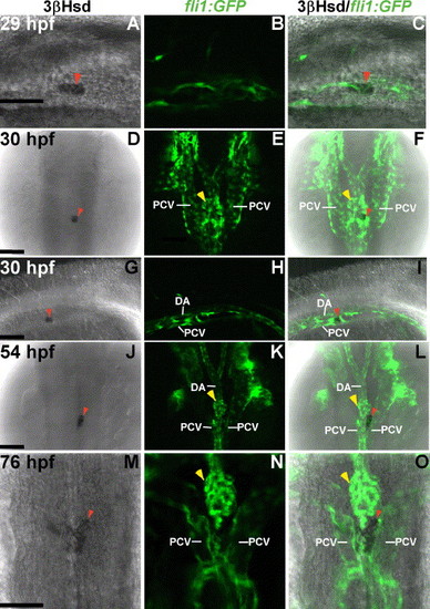
Interaction of interrenal and endothelial cells as revealed in Tg(fli1:EGFP)Y1 transgenic zebrafish. Single confocal sections showing the interrenal tissues as stained by 3β-Hsd activity assay (left panel: A, D, G, J, M), and the neighboring ECs as labeled by green fluorescence (middle panel: B, E, H, K, N), of Tg(fli1:EGFP)Y1 embryos while staged at 29 hpf (A–C), 30 hpf (D–I), 54 hpf (J–L) and 76 hpf (M–O), respectively. The merged images of 3β-Hsd activity and GFP are shown in the right panel (C, F, I, L, O). Panels A–C and panels G–I are lateral views with anterior oriented to the right. Panels D–F and panels J–O are dorsal views with anterior oriented to the top. The interrenal cells are in close contact with ECs that are engaged in axial vessel assembly. DA, dorsal aorta; PCV, posterior cardinal vein. Red arrowheads indicate interrenal tissues, while yellow arrowheads indicate glomeruli. Scale bar, 50 μM. |
| Gene: | |
|---|---|
| Fish: | |
| Anatomical Term: | |
| Stage Range: | Prim-5 to Protruding-mouth |
Reprinted from Developmental Biology, 297(1), Liu, Y.W., and Guo, L., Endothelium is required for the promotion of interrenal morphogenetic movement during early zebrafish development, 44-58, Copyright (2006) with permission from Elsevier. Full text @ Dev. Biol.