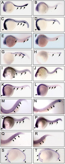
Expression of hematopoietic and vascular markers in wild type and zbp-89 morphants. (A-R) Expression of primitive erythroid (A-D), primitive myeloid (E-J) and definitive hematopoietic markers (K-R). Normal expression (arrows) of the primitive erythroid genes gata1 (A) and tif1g (C) in the anterior ICM of wild-type embryos is almost completely lost in zbp-89 morphants (B,D, respectively). Expression of tif1g in neural tissue (arrowheads) is not affected. Expression of the primitive myeloid markers pu.1, mpo and l-plastin in wild type (E,G,I) and in zbp-89 morphants (F,H,J). The normal expression of pu.1 in primitive macrophages in the anterior ICM (arrow) in 24 hpf embryos (E) is markedly reduced by depletion of ZBP-89 (F). Its expression is also reduced in 20 hpf embryos in the head, rostral blood islands and ICM (not shown). mpo (G) and l-plastin (I) are normally expressed in the ICM of 24 hpf embryos (arrows) and in the anterior yolk region (l-plastin). Both markers are severely reduced by the loss of ZBP-89 (H,J, respectively). (K-R) Expression of the definitive hematopoietic markers runx1 and c-myb. Expression of runx1 begins in the ICM (K, arrows) at 24 hpf and is well developed in the ventral dorsal aorta at 48 hpf (M, arrows). Loss of ZBP-89 markedly reduces expression of runx1 in 24 hpf (L) and 48 hpf (N) embryos. c-myb is normally expressed in the ICM of wild-type embryos at 24 hpf (O, arrows). In 48 hpf embryos, cells expressing c-myb are found scattered along the ventral wall of the dorsal aorta (Q, arrows), within the first progenitors of definitive hematopoiesis. c-myb expression is significantly reduced in zbp-89 morphants in both 24 and 48 hpf embryos (P,R). Non-hematopoietic expression of runx1 (K,L) and c-myb (O,P) in neural tissue (arrowheads) was not affected by the loss of ZBP-89. (S,T) flk1 expression in 20 hpf wild type and in zbp-89 morphants. flk1 is normally expressed in cells located in two strips of the anterior lateral mesoderm (short arrows), and in the forming anterior (arrows) and posterior (arrowheads) ICM (S). This expression was not affected by the loss of ZBP-89 (T). All views are lateral with anterior left and dorsal top.
|
