
Fig. 1
- ID
- ZDB-FIG-060808-231
- Publication
- Stock et al., 2006 - Developmental genetic mechanisms of evolutionary tooth loss in cypriniform fishes
- Other Figures
- All Figure Page
- Back to All Figure Page
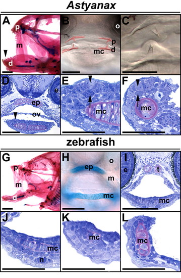
Oral morphology in Astyanax and zebrafish. (A) Teeth are present on premaxillary (p), maxillary (m) and dentary bones (d, arrowhead) of adult Astyanax (lateral view, cleared and stained with Alcian blue and Alizarin red). (B,C) A single tooth is present on each premaxillary and dentary bone in 120 hpf Astyanax (frontal views; teeth and bones digitally colored red in B). (D-F) Bell-shaped tooth germs in 72 hpf Astyanax lower jaw. Dental epithelium indicated by arrowhead, darkly-stained dental mesenchyme by arrow. Transverse sections in D,E; sagittal in F. (G) Toothless oral cavity of adult zebrafish. (H) Toothless oral cavity of 124 hpf zebrafish larva cleared and stained with Alcian green. (I-L) No tooth germs are visible in sectioned, Toluidine blue-stained zebrafish larvae. (I,J) Identical transverse sections of a 72 hpf specimen. (K,L) Sagittal views of the lower jaw of 72 hpf and 120 hpf specimens, respectively. d, dentary; e, eye; ep, ethmoid plate; m, maxillary; mc, Meckel's cartilage; n, neuromast; o, olfactory organ; ov, oral valve; p, premaxillary; t, trabecula. Scale bars: 1 mm in A,G; 100 μm in B,D,H,I; 50 μm in C,E,F,J-L. |