
Fig. S1
- ID
- ZDB-FIG-060710-9
- Publication
- Muto et al., 2006 - Rab11-FIP4 is predominantly expressed in neural tissues and involved in proliferation as well as in differentiation during zebrafish retinal development
- Other Figures
- All Figure Page
- Back to All Figure Page
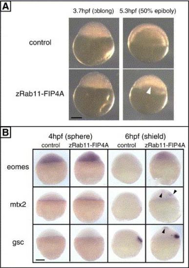
Supplementary Fig. 1. Functional analysis of zRab11-FIP4 by ectopic expression. (A) Morphology of early embryos injected with EGFP mRNA alone (upper) or in combination with mRNA for zRab11-FIP4A (lower) is shown. Three hundred picograms each of RNAs was injected. Animal pole is at the top. Morphological defects in the zRab11-FIP4A-expressing embryos were not observed at oblong stage (left, 3.7 hpf) but appeared after the onset of epiboly (right, 5.3 hpf) as an unequal distribution of deep cells (white arrowhead). Most of these embryos then underwent lysis between 8 and 12 hpf (see also Table 1). (B) Effect of ectopically expressed zRab11-FIP4A on gene expression. The expression of eomesodermin (eomes), mtx2 and goosecoid (gsc) before (4 hpf) and after (6 hpf) the appearance of the morphological phenotype. Positions of ectopically expressed of mtx2 and gsc are indicated by black arrowheads. The animal pole is at the top, and scale bar indicates 200 µm. |
| Genes: | |
|---|---|
| Fish: | |
| Anatomical Terms: | |
| Stage Range: | Sphere to Shield |
Reprinted from Developmental Biology, 292(1), Muto, A., Arai, K.I., and Watanabe, S., Rab11-FIP4 is predominantly expressed in neural tissues and involved in proliferation as well as in differentiation during zebrafish retinal development, 90-102, Copyright (2006) with permission from Elsevier. Full text @ Dev. Biol.