
Fig. 7
- ID
- ZDB-FIG-060628-30
- Publication
- Wei et al., 2004 - The zebrafish Pard3 ortholog is required for separation of the eye fields and retinal lamination
- Other Figures
- All Figure Page
- Back to All Figure Page
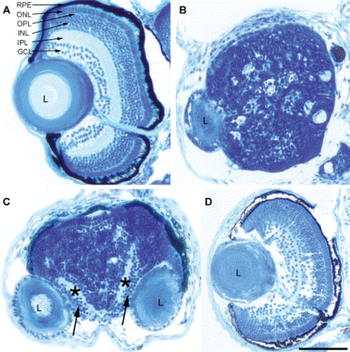
Loss of Pard3 function disrupted retinal patterning. At 5 dpf, histology was performed on a wild-type embryo (A), an anti-pard3 morpholino-injected cyclopic embryo (B), an anti-pard3 morpholino-injected embryo that developed fused retinas with separated lenses (C), and an embryo that was co-injected with anti-pard3 Morph-1 and in vitro-transcribed pard3 mRNA (encoding the 150-kDa Pard3 open reading frame flanked by the β-globin 5′ and 3′ UTRs; D). The ganglion cell and inner plexiform layers are marked with arrows and asterisks, respectively (C). RPE, retinal pigmented epithelium; ONL, outer nuclear layer; OPL, outer plexiform layer; INL, inner nuclear layer; IPL, inner plexiform layer; GCL, ganglion cell layer; L, lens. Scale bar indicates 100 μm. |
| Fish: | |
|---|---|
| Knockdown Reagent: | |
| Observed In: | |
| Stage: | Day 5 |
Reprinted from Developmental Biology, 269(1), Wei, X., Cheng, Y., Luo, Y., Shi, X., Nelson, S., and Hyde, D.R., The zebrafish Pard3 ortholog is required for separation of the eye fields and retinal lamination, 286-301, Copyright (2004) with permission from Elsevier. Full text @ Dev. Biol.