|
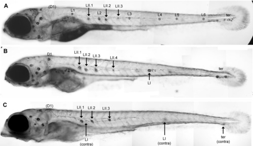
Effect of tacstd inactivation on secondary lateral line formation. A: A normal 6-day-old larva showing three primII neuromasts (LII.1, LII.2, and LII.3). B,C: Two morphant embryos showing a strong phenotype. B: One lateral and two terminal primI neuromasts are present. The primII neuromasts have formed normally (LII.1-LII.4). C: No primI neuromast is present on the focused side of the embryo; two lateral and one terminal primI neuromasts are present on the other side as indicated. This embryo also shows the stereotyped head defect present in about 30% of the embryos. Note that in A and C, the D1 neuromast has been dislodged during manipulation. This rarely happens and can easily be detected because a rim of labelled cells remains attached to the epidermis, as can be faintly seen at this low magnification in A.
|