
Fig. 6
- ID
- ZDB-FIG-060605-28
- Publication
- Pyati et al., 2006 - Sustained Bmp signaling is essential for cloaca development in zebrafish
- Other Figures
- All Figure Page
- Back to All Figure Page
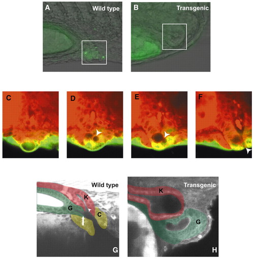
Visualizing cloaca development in living embryos. (A,B) Acridine Orange staining in wild-type and transgenic embryos to assay cell death in the developing cloaca region. The posterior kidney region is boxed. (C-F) Detailed time lapse of an msxb-gfp embryo during opening of the presumptive cloaca. Note initially the kidney terminus, cup-shaped proctodeum, and epidermis (green; C) at 24-somites. A single vacuolated proctodeal cell (arrowhead in D-F) emerges and migrates to the ventral limit of the epidermis, where it forms a pore (F). At that point, the kidney terminus has connected to the epidermis and there is a continuous opening to the outside of the embryo (see also Movie 1 in the supplementary material for the full time lapse). (G,H) Excretory region of a wild-type and transgenic sibling larva, respectively, at 4 dpf. Regions of the excretory system have been pseudo-colored for identification: K, kidney (red); G, gut (green); C, cloaca (yellow). Arrowhead in G marks the kidney (urogenital) opening, and arrow marks the gut opening. |