
Fig. 7
- ID
- ZDB-FIG-060522-21
- Publication
- Wang et al., 2006 - Dishevelled genes mediate a conserved mammalian PCP pathway to regulate convergent extension during neurulation
- Other Figures
- All Figure Page
- Back to All Figure Page
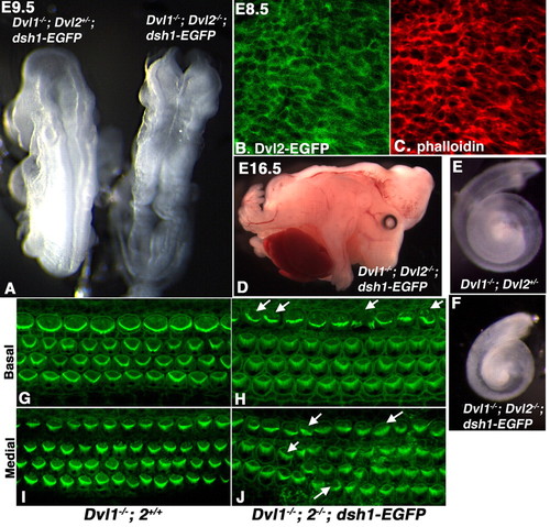
dsh1-EGFP failed to rescue the craniorachischisis, cochlear elongation and stereociliary polarity phenotypes in Dvl1-/-; Dvl2-/- embryos. (A,D) Dvl1-/-; Dvl2-/-; dsh1-EGFP embryos (right in A) displayed craniorachischisis at E9.5 and E16.5, suggesting that dsh1-EGFP was not able to compensate for the convergent extension defect in neurulation Dvl1-/-; Dvl2-/- mutants. (B) Confocal analysis indicated that dsh1-EGFP was primarily localized to the plasma membrane in the neuroepithelium. (C) Confocal analysis of phalloidin staining. (E,F) Cochlea recovered at p0 from Dvl1-/-; Dvl2+/- and Dvl1-/-; Dvl2-/-; dsh1-EGFP mice, indicating that dsh1-EGFP also failed to rescue the cochlea elongation defect in Dvl1-/-; Dvl2-/- mutants that was also thought to be due to disruption of convergent extension. (G,I) Confocal scan at either the base (G) or medial region (I) showing uniform stereociliary orientation in the organ of Corti of a p0 Dvl1-/-; Dvl2+/+ mouse. (H,J) However, in the organ of Corti of a Dvl1-/-; Dvl2-/-; dsh1-EGFP mouse, the stereociliary bundles were mis-oriented (arrows) in some hair cells. Misalignment in the outer hair cells could also be observed. |