
Fig. 3
- ID
- ZDB-FIG-060328-10
- Publication
- Sumanas et al., 2006 - Ets1-Related Protein Is a Key Regulator of Vasculogenesis in Zebrafish
- Other Figures
- All Figure Page
- Back to All Figure Page
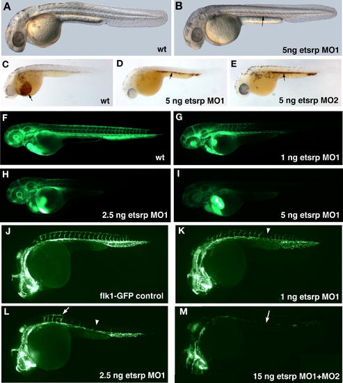
MO Knockdown of Etsrp Protein Function Disrupts Blood Vessel Formation in the Zebrafish Embryos (A,B) Morphological analysis of live etsrp morphants at 34 hpf. (A) Uninjected control embryo. (B) 5 ng of etsrp MO1-injected embryo. Notice that red blood cells are scattered throughout the circulatory system in the control uninjected embryo while they accumulate at their formation site within the intermediate cell mass (arrow, B) in the etsrp morphant. (C–E) o-dianisidine staining of heme in the red blood cells of uninjected control (C), 5 ng of etsrp MO1-injected (D) and 5 ng of etsrp MO2-injected (E) embryos at 34 hpf. While many circulating blood cells are apparent within the common cardinal vein before entering the heart in the control embryo (arrow, C), they stay at their formation site within the ICM region in etsrp morphants (arrows). (F–I) Microangiography analysis of the circulatory system by injecting fluorescein-labeled dextran into the sinus venosus of etsrp morphants at 55 hpf. (F) Control uninjected, (G) 1 ng of etsrp MO1-injected (H) 2.5 ng of etsrp MO1-injected (I) 5 ng of etsrp MO1-injected embryos. Note that the embryo in (G) has lost circulation in the posterior vessels, the embryo in (H) has lost circulation in most vessels, and the embryo in (I) has no circulation at all. (J–M) Analysis of blood vessels in live flk1-GFP transgenic embryos at 26 hpf. (J) Control uninjected, (K) 1 ng of etsrp MO1-injected, (L) 2.5 ng of etsrp MO1-injected, (M) 15 ng of etsrp MO1+MO2 (1:1) mix-injected embryos. Note the gaps in formation of intersegmental vessels in (K) (arrowhead), the missing (arrowhead) and abnormally branched, (arrow) intersegmental vessels in (L), and the nearly completely eliminated flk1 expression from axial vessels (arrow) in (M). |