
FIGURE
Fig. 2
- ID
- ZDB-FIG-060303-6
- Publication
- Milan et al., 2006 - Notch1b and neuregulin are required for specification of central cardiac conduction tissue
- Other Figures
- All Figure Page
- Back to All Figure Page
Fig. 2
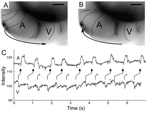
Characteristics of retrograde conduction in the zebrafish heart at 48 hpf. (A,B) Calcium activation maps for normal anterograde cardiac cycle (A) and spontaneous premature ventricular beat (B). Both anterograde and retrograde AV delay are evident as compressed spacing of isochronal lines (100 msecond intervals). Scale bars: 50 μm. (C) Contemporaneous recordings of atrial and ventricular contraction using intensity-time plots from regions over the respective chambers, during ventricular pacing at 48 hpf. The ladder diagram depicts the resulting Wenckebach-type retrograde ventriculo-atrial block. |
Expression Data
Expression Detail
Antibody Labeling
Phenotype Data
Phenotype Detail
Acknowledgments
This image is the copyrighted work of the attributed author or publisher, and
ZFIN has permission only to display this image to its users.
Additional permissions should be obtained from the applicable author or publisher of the image.
Full text @ Development