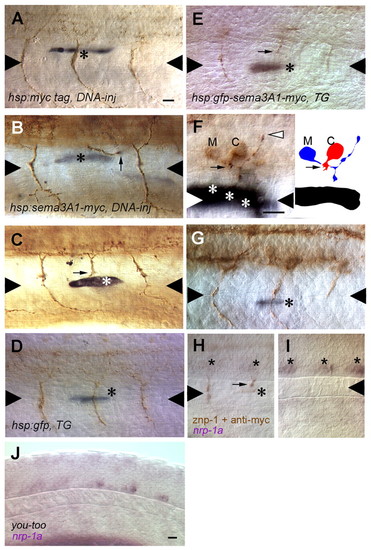
CaP axons are repulsed by myotome cells that express ectopic Sema3a1 along the common pathway but not by those along the CaP specific ventral pathway. (A) A CaP axon is not perturbed by a myotome cell (asterisk) along the common pathway that expresses the Myc epitope following heat induction of a hsp70:myc injected embryo. Triangles denote the horizontal myoseptum. (B) A CaP axon (arrow) appears to have turned away to avoid a myotome cell (asterisk) along the common pathway that expresses ectopic Sema3a1 following heat induction of a hsp70:sema3a1-myc-injected embryo. CaP axons in the segment anterior and posterior to the repulsed CaP follow a normal trajectory. The myotome cell expressing ectopic Sema3a1 in the posterior segment is out of the focal plane of the CaP axon and is located lateral to the medial myotome cells lining the common pathway. (C) A CaP axon (arrow) appears to have stalled when encountering a myotome cell (asterisk) along the common pathway that expresses ectopic Sema3a1 following heat induction of a hsp70: sema3a1-myc-injected embryo. (D) A CaP axon is not perturbed by a muscle pioneer cell (asterisk) laser induced to express GFP in a hsp70:gfp transgenic embryo. (E) A CaP axon (arrow) is stalled in the vicinity of a muscle pioneer (asterisk) laser induced to express ectopic Sema3a1 in a hsp70:gfp-sema3a1-myc transgenic embryo. (F) A CaP axon (arrow) is stalled in the vicinity of three myotome cells (asterisks) along the common pathway laser induced to express ectopic Sema3a1 in a hsp70:gfp-sema3a1-myc transgenic embryo, but a presumptive MiP axon (white arrowhead) is normal. Right panel shows a camera lucida drawing of the CaP and MiP motoneurons. C, the CaP cell body; M, the MiP cell body. (G) A CaP axon appears normal despite encountering a ventral muscle fiber beyond the horizontal myoseptum laser-induce to express ectopic Sema3a1 in a hsp70:gfp-sema3a1-myc transgenic embryo. The CaP axon immediately posterior to the experimental CaP axon maybe shorter because it is paused at the choice point. (H) nrp1a expression is downregulated in CaP neurons, despite not having reached the horizontal myoseptum because of stalling in the vicinity of a myotome cell (asterisk) laser induced to express ectopic Sema3a1 in a hsp70:gfp-sema3a1-myc transgenic embryo. Stars indicate CaP cell bodies. (I) CaPs (stars) in more caudal segments in the embryo shown in H have not yet projected axons but do express nrp1a. (J) nrp1a is downregulated in CaP neurons in you-too mutants (26 somite-stage; 22 hpf) in which CaP axons fail to extend properly along the common pathway. Scale bars: 20 μm.
|
