
Fig. 3
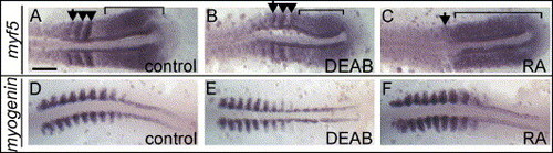
Regulation of early and late differentiation markers by RA. 10-somite stage embryos are displayed as in Fig. 2. (A) myf5 is expressed in wild-type posterior psm (bracket) and is re-expressed in forming somites (arrowheads). Somitic expression decreases as somites mature (arrow indicates the posterior border of last formed somite). (B) In DEAB-treated embryos, myf5 is reduced in both psm (bracket) and somites (arrowheads). (C) In RA-treated embryos, myf5 expression encompasses the entire psm (bracket; arrow indicates the posterior border of last formed somite) and is absent in somites. (D) myogenin expression in wild-type embryos resembles that of myoD, it is decreased in posterior somites in DEAB-treated embryo (E) and increased in somites of RA-treated embryo (F). Scale bar: in panel A, 100 μm for panels A–F. |
| Genes: | |
|---|---|
| Fish: | |
| Conditions: | |
| Anatomical Terms: | |
| Stage: | 10-13 somites |
Reprinted from Developmental Biology, 289(1), Hamade, A., Deries, M., Begemann, G., Bally-Cuif, L., Genet, C., Sabatier, F., Bonnieu, A., and Cousin, X., Retinoic acid activates myogenesis in vivo through Fgf8 signalling, 127-140, Copyright (2006) with permission from Elsevier. Full text @ Dev. Biol.