|
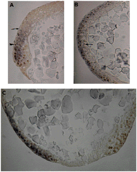
Sections of embryos stained for eve1 and Ntl. The eve1 staining appears as a dark purple cellular coloration after wholemount in situ hybridizations using the chromogenic alkaline phosphatase reaction, while antibody stainings for Ntl give a brown color in nuclei subsequent to a peroxidase detection. (A) Sagittal section through a germ-ring stage embryo. The extents of eve1 or Ntl staining are indicated by an arrow and an arrowhead, respectively. (B) Ventral region of a shield stage embryo. Equatorial section. Epiblast is positive for eve1 and Ntl. In hypoblast, there is no eve1 staining. One nucleus, faintly stained for Ntl, is indicated by an arrow. (C) Sagittal section through the vegetal region of a 90% epiboly embryo. Ntl staining is on both sides of yolk plug, eve1 staining is ventral, on the left side.
|