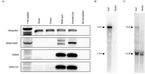
Zebrafish zonadhesin mRNA expression. A) Semi-quantitative reverse transcription PCR analysis of zebrafish tissues. Three primer sets were designed against zonadhesin specific sites. The amplicons of the first primer set crossed the mucin domain stretching from the MAM domains to the first VWD domain (MAM-VWD). The second primer set amplified within the VWD9 domain (VWD9). Products of the third primer set stretched from the last VWD domain to the cytoplasmic domain (VWD9-CD). Ubiquitin primers were used as a positive control and a template-free reaction was included as a negative control. B-C) Expression of zebrafish zonadhesin analyzed by Northern blot. B) Expression of zonadhesin was investigated in the zebrafish testis and gut. Five micrograms of total RNA from each tissue was blotted on a positively charged nylon membrane and hybridized with a DNA probe encompassing the zebrafish EGF to CD region of zan. C) The same Northern blot membrane was reprobed with zebrafish alpha-tubulin as a positive control.
|
