
Fig. 2
- ID
- ZDB-FIG-051220-22
- Publication
- Otteson et al., 2005 - Genomic organization of zebrafish cone-rod homeobox gene and exclusion as a candidate gene for retinal degeneration in niezerka and mikre oko
- Other Figures
- All Figure Page
- Back to All Figure Page
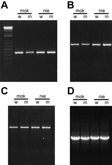
Photographs of ethidium bromide-stained agarose gels showing representative PCR amplifications of zebrafish crx from genomic DNA isolated from mok (mikre oko), nie (niezerka) homozygous mutants (m) and their phenotypically wild-type sibs (w) using primers listed in Table 1. A: 5' end of Exon 1 including repetitive region within 5' untranslated region (UTR). B: 5' coding region of Exon 3 and flanking portion of Intron 2. C: 3' end of coding region of exon 3 and repetitive sequences in 3' UTR. D: Intron 2 shows a small, but detectable difference in the size between mok (1026 bp) and nie (1064 bp) although within each line, the amplicons from homozygous mutant and wild-type sibs co-migrate. |