|
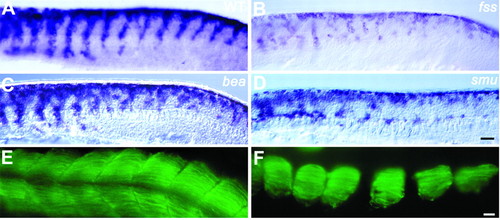
In segmentation mutants, neural crest migration is disrupted in the same somites in which segmentation is disrupted. Whole-mount staining with a crestin riboprobe of wild-type (A) fss (B), bea (C) and smu mutants (D) at 21 hpf to reveal neural crest cells. (A) In wild-type embryos, neural crest cells migrate in segmental streams along the entire AP axis. (B) Neural crest migration is abnormal from the first somite in fss mutants. (C) In bea mutants, neural crest migration is disrupted posterior to somite 6. (D) In smu mutants there is no clear segmental pattern of neural crest migration; in addition, neural crest cells stall at the level of the dorsal aspect of the notochord. (E,F) EB165 antibody staining reveals that in smu mutants (F) somites are smaller along the DV axis, but a significant amount of fast muscle is still present, although there may be less than in wild types (E). Scale bar: 60 µm in A-D; 25 µm in E,F.
|