
Fig. 7
- ID
- ZDB-FIG-050916-7
- Publication
- Beis et al., 2005 - Genetic and cellular analyses of zebrafish atrioventricular cushion and valve development
- Other Figures
- All Figure Page
- Back to All Figure Page
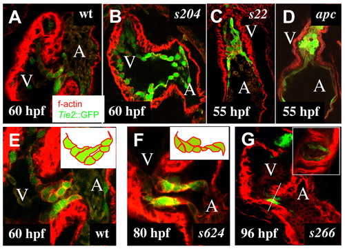
AV canal defective mutants identified in a large-scale mutagenesis screen. Wild-type (A,E) and mutant (B-D,F,G) hearts between 55 and 96 hpf. Confocal images of embryonic hearts at 60 (A,B) and 55 hpf (C,D). Confocal images of the AV canal at 60 (E), 80 (F) and 96 hpf (G). In contrast to wild-type hearts at 60 hpf (A), in s204 mutant hearts (B), the AV canal endocardial cells fail to adopt a cuboidal shape. (C) In s22 mutant hearts, the ventricular lumen contains cuboidal endocardial cells. (D) In the apc mutant heart, the ventricular lumen is filled with Tg(Tie2:EGFP)s849-positive cells with a mesenchymal morphology. (E) In wild-type embryos, only AV canal endocardial cells located next to the ventricular border form cellular extensions into the AV canal ECM and extensions are directed towards the atrial border of the AV canal. (F) In s624 mutant embryos at 80 hpf, no ECs have formed, and the AV canal endocardial cells extend cellular projections in different directions. (G) In s266 mutant embryos, no ECs have formed by 96 hpf; the AV canal is lined by a single layer of cuboidal cells. Inset shows a transverse section of the s266 mutant AV canal (compare with Fig. 2F and see Movie 2 in the supplementary material). A, atrium; V, ventricle.
|