
Fig. 5
- ID
- ZDB-FIG-050913-5
- Publication
- Wada et al., 2005 - Hedgehog signaling is required for cranial neural crest morphogenesis and chondrogenesis at the midline in the zebrafish skull
- Other Figures
- All Figure Page
- Back to All Figure Page
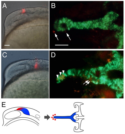
Fate maps in Hh-deficient embryos. (A,C) PKH labeling immediately after injection, lateral views. (B, D) Labeled cells at 80 hpf derived from these injections, showing co-localization (yellow) of PKH (red) and sox10:egfp (green) in dissected preparations, ventral views. (A) NC cells labeled dorsal to the optic vesicle do not contribute to cartilage, but remain undifferentiated (B, arrows). (C) By contrast, cells labeled posterior to the optic vesicle contribute to the midline cartilage rod (D, arrowheads) and posterior trabeculae (arrows). (E) Schematic representation of the fate map in Hh-deficient embryos. Trabecular precursors form cartilage (blue), whereas ethmoid progenitors (red) do not. Scale bars: 50μm; in A for A,C; in B for B,D. |