|
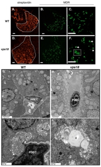
Mutation in vps18 results in biliary defects. (A-F) Confocal images of livers from wild-type (top) and vps18 mutant (bottom) embryos from day 5 (A,B,D,E) and day 7 (C,F) labeled with CY3-SA (A,D) and anti-MDR (B,C,E,F). Some hepatocytes in the mutant embryos retain some MDR in the cytoplasm (box in F). Scale bars: 10 µm. (G-I) TEM of day 5 embryos identifies PDECs as cells adjacent to hepatocytes that collect bile from the canaliculi of neighboring hepatocytes and transport it through their lumen (arrows in I). (H,J) vps18 mutant livers contain virtually no PDECs, and those that are found do not establish contacts with hepatocytes, are shrunken and have condensed DNA (H). PDEC, pre-ductal epithelial cell; c, canaliculi; c?, putative aberrant canaliculi; L, lumen. Scale bars: 20 µm G,H; 500 nm in I,J.
|