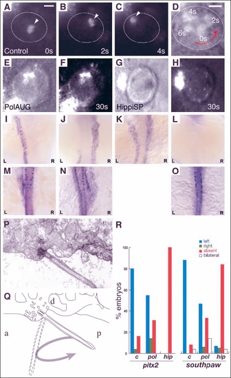
Impaired fluid flow in Kupffer's vesicle is associated with defects in laterality. Embryos at the 8-10 somite stage were dechorionated and fluorescent beads were injected into Kupffer's vesicle. Control embryos showed a rotating movement of bead aggregates in a counterclockwise orientation (arrowheads in A-C) when viewed dorsally. Relative timepoints in seconds are indicated in the bottom right of each panel. The wall of Kupffer's vesicle is indicated by dotted lines. (D) Superposition and enlargement of frames (A-C) with an additional transmitted light frame showing the counterclockwise direction of movement. None of the injected morphant embryos (polarisAUG and hippiSP) showed this phenomenon (E-H). Abnormal expression of the laterality markers pitx2 and spaw in polarisAUG and hippiSP embryos (I-O,R). In situ experiments were performed on 14-somite (spaw) and 20-somite (pitx2) embryos. Dorsal views of the lateral plate mesoderm are shown with the different expression patterns seen in polarisAUG embryos. southpaw was expressed on the left (I), right (J) or bilaterally (K), or in many cases absent (L). pitx2 shows the same patterns (M, left-sided; N, right-sided; O, absent expression), with the exception of bilateral expression. Sagittal section electron micrograph (P) of the roof of Kupffer's vesicle showing, a single cilium and associated basal body. (Q) Diagram of the micrograph in P, detailing how the angle of the basal body [approximately 45° to the posterior (P)] would result in the cilium projecting into Kupffer's vesicle on the right-to-left portion of the counterclockwise rotary beat. (R) Frequency of laterality defects in polaris and hippi morphant embryos. Expression of pitx2 and southpaw was randomized in polarisAUG embryos, while hippiSP embryos showed significantly higher numbers of embryos with no expression of southpaw and pitx2 (control, n=25; polarisAUG/pitx2, n=29; polarisAUG/southpaw, n=36; hippiSP/pitx2, n=40; hippiSP/southpaw, n=57). In embryos lacking laterality signals in the lateral plate mesoderm, gene expression was nevertheless maintained in the tailbud (spaw) and Rohon-Beard neurons (pitx2). Scale bar: 10 µm. a, anterior; d, dorsal.
|
