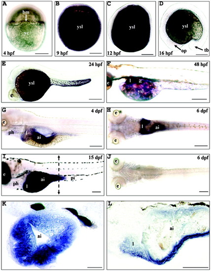
Expression pattern of the microsomal triglyceride transfer protein (MTP) large subunit gene (mtp) during zebrafish embryonic and larval development. A: After the mid-blastula transition, 4 hours postfertilization (hpf). B: During the gastrulation period (9 hpf). C: At the five-somite stage (12 hpf). D: At the 15-somite stage (16 hpf). E: A 24-hpf embryo. F: A 48-hpf embryo. G: A 4 days postfertilization (dpf) larva. H,J: A 6 dpf larva. I,K,L: A15 dpf larva. Whole-mount in situ hybridizations were performed with digoxigenin-labeled sense and antisense riboprobes and are presented in lateral (A-G,I) or dorsal (H,J) views with the anterior part to the left (C-J,L). The 6-dpf and 15-dpf larvae were nourished ad libitum. In some cases (G,H,J), the animals were raised in 0.2 mM 1-phenyl-2-thio-urea containing water to prevent pigment formation. The hybridization signal is colored dark brown to blue. J: No staining signal was observed by using the sense probe. A: A specific hybridization signal was first observed in the blastoderm margin (bm). B-F: mtp was then strongly expressed in the yolk syncytial layer (ysl) from the end of the gastrulation period until 48 hpf. C-E: No transcripts were detected in embryonic structures until 24 hpf. F: At 48 hpf, mtp mRNA was detected in the liver (l), intestinal tube (it), and yolk syncytial layer. G: By 4 dpf, the hybridization signal was restricted to the liver and anterior intestine (ai). H: By 6 dpf, the two liver lobes and the anterior intestine were strongly labeled. I: By 15 dpf, mtp mRNA was detected in the liver and the anterior intestine, including the intestinal bulb, while no hybridization signal could be detected in pharynx (ph) and posterior intestine (pi). Transverse (K), indicated by a vertical broken line in (I), and parasagittal (L) cryosections of 15-dpf larvae showed that mtp transcripts were restricted to enterocytes and hepatic cells. Embryos, larvae, and cryosections were mounted in 100% glycerol. yc, yolk cell; op, optic primordium; tb, tail bud; e, eye. Scale bars = 100 μm in A-D,F-L, 500 μm in E.
|
