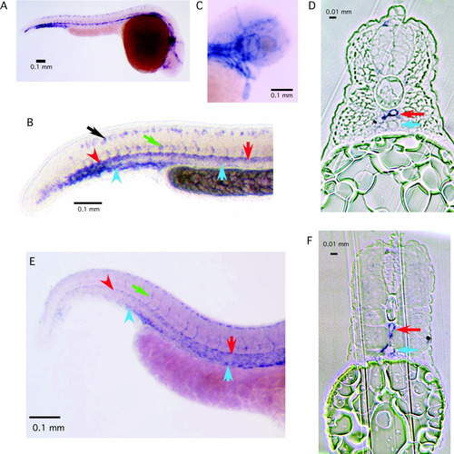
Whole-mount in situ hybridization of CD146 in zebrafish. The digoxigenin-labeled antisense probe was derived from the first 0.6 kb of the mRNA. A: Overall staining pattern of CD146 in a 24-hour postfertilization (hpf) embryo. B: A magnification at the trunk-tail region of a 24-hpf embryo. Notice the staining of the intersomitic vessels (green arrow), dorsal aorta (red arrow), caudal artery (red arrowhead), and the caudal vein (blue arrowhead). Staining of the posterior cardinal vein was noticeably absent at this stage (blue arrow). Staining of neuronal cells was also seen at this stage (black arrow). C: Staining pattern in the head region of an embryo at 3 days postfertilization. D: Cross-section of an embryo at 24 hpf at the trunk region showing the expression of CD146 in the artery and a very low level of expression in the vein. E: Expression pattern of CD146 mRNA in a 2-day-old embryo showing endothelial specificity. Notice the expression of CD146 in the posterior cardinal vein. F: Cross-section of a 3-day-old embryo at the trunk region showing expression of CD146 in both the dorsal aorta and posterior cardinal vein.
|
